Neisser Staining
Staining according to Neisser is a test for the presence of polyphosphates stored in the cells (= storage materials). This method is an indispensable aid to the identification of certain strains of filamentous bacteria. Furthermore, this staining method can make the Bio-P bacteria, responsible for biological phosphate removal, visible.
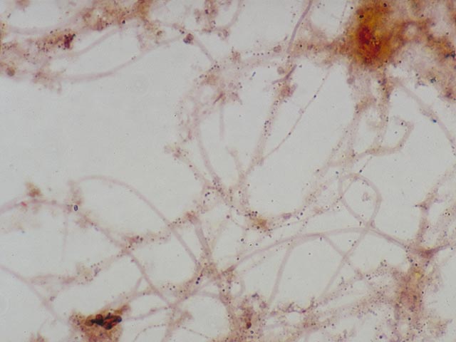
Neisser negative cells stain hardly or not at all (slightly brown or yellow; Three main groups of Neisser positive bacteria can be distinguished.
1. Filamentous bacteria which stain completely grey-violet . This almost always applies to Nostocoida limicola or Type 0092.
2. Filamentous bacteria which contain blue-black coloured polyphosphate globules . Without staining, these globules cannot be clearly observed with a light microscope. They are indeed clearly visible if a much higher magnification (electron microscopy) is used . These globules, which are present in pairs, are an important identification characteristic for Microthrix parvicella.
3. Colonies of blue-black coloured cells . These are comprised of Bio-P bacteria. There are some variations in the manner in which these types of colonies stain with Neisser. The shade is sometimes much lighter , or only a part of the cell stains darkly.
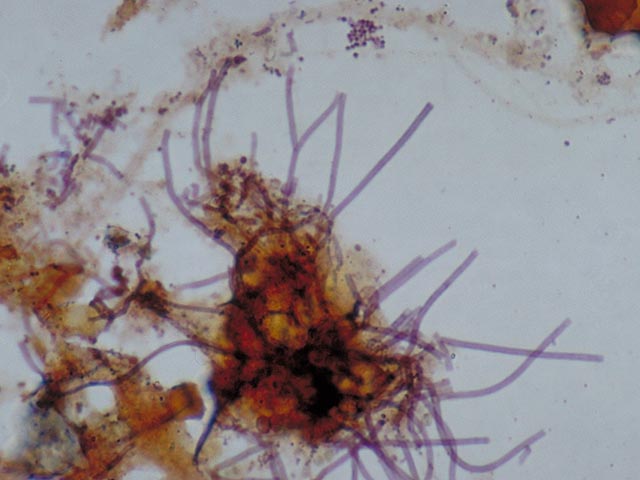
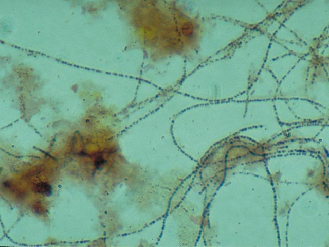
Neisser – Negative Neisser – Positive Neisser – Positive